Applications in Oncology Research
Examples of Efficacy Testing of Anti-Cancer Drugs
Design of Cancer / Stromal Co-Culture Model
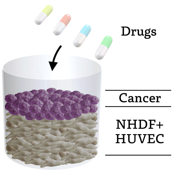
Schematic image of invivoid® cancer / stromal co-culture model
*NHDF: Normal Human Dermal Fibroblasts
*HUVEC: Human Umbilical Vein Endothelial Cells
Capillary Blood Vessel Network
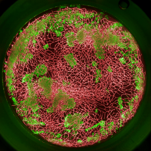
Example HCA image of cancer / stromal co-culture model. Patient-derived cancer cells are growing on the stromal layer containing capillary blood vessel network.
*green: anti-EpCAM antibody (Patient-derived CRC cells)
*red: anti-CD31 antibody (Vascular Endothelial Cells)
Cancer Cell Invasion into Blood Vessels
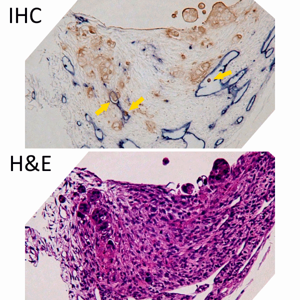
Example of section image of cancer / stromal co-culture model. Invasion of cancer cells into blood vessels (yellow arrows) were observed.
--IHC--
*blue: anti-CD31 antibody (Vascular Endothelial Cells)
*brown: anti-CEA antibody (Patient-derived CRC Cells)
Efficacy Testing
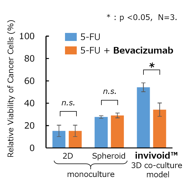
Representative result of cancer cell viability testing. Relative viability of cancer cells were evaluated. Combination effect of 5-FU and Bevacizumab was observed only in invivoid® 3D co-culture model.
(Colorectal cancer cell line: HCT 116)
Examples of Efficacy Evaluation of Immuno-Oncology Agents
Design of Cancer / Stromal Co-Culture Models for Evaluating I-O Agents
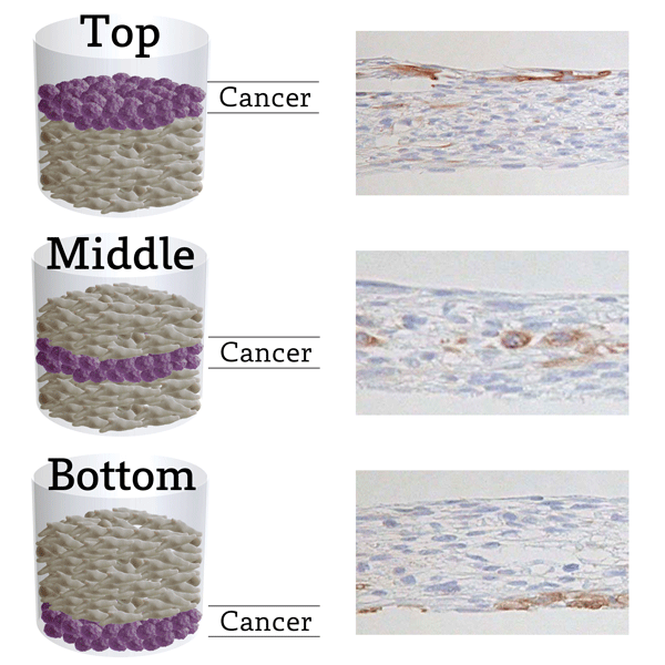
3D co-culture models designed to mimic "stromal barrier" that blocks infiltration of immune cells. Cancer cells can be placed at a desired position in the tissue.
*blue: hematoxylin
*brown: anti-cytokeratin 7 antibody (Lung cancer cells)
Evaluation Scheme of I-O Agents
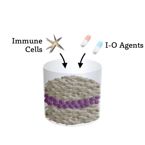
1. Construct cancer / stromal co-culture model and culture for several days
2. Add immune cells and I-O agents
3. Evaluate viability of cancer cells and infiltration of immune cells, etc.
Cancer Cell Killing Assay
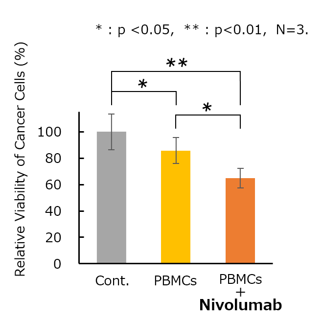
Representative result of cancer cell killing assay (allogeneic model). Significant difference between "PBMCs" and "PBMCs+Nivolumab" was observed.
(Lung cancer cell line: NCI-H1975)
*PBMCs: Peripheral Blood Mononuclear Cells
Virtual TILs Evaluation
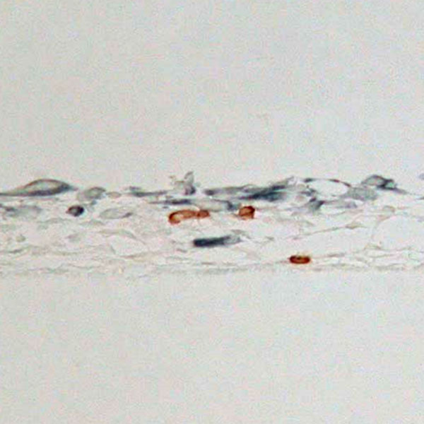
Example of section image (Immunohistochemistry: IHC). Cytotoxic T-cells (brown) which had infiltrated into the tissue were observed.
*blue: anti-cytokeratin 7 antibody (Lung cancer cells)
*brown: anti-CD8 antibody (Cytotoxic T-cells)
*TILs:Tumor-Infiltrating Lymphocytes